 企业邮箱
企业邮箱


Discovery of human secondary life cells and regenerative nutrient substances for fulfilling organ life relay

The keynote speech on the 9th Chinese National Academic Conference of Burns, Wounds and Ulcers
In my speech, I would like to address our exciting achievements in regenerative medical research and the journey from burn research to exploration of life mysteries; from discovery of potential regenerative cells, or the secondary life cells to discovery of regenerative nutrient substances, or the source of regenerative energy. Based on these discoveries, we have made a series of extraordinary inventions which can be utilized to carry out in situ organ regeneration for human organ life relay. Before I introduce our accomplishments and their applications in life science, let's take a look of the history in human life science research.
I. Life Science Study Found Hope in Struggles
It has been more than 160 years since the start of cytological study in 1840. During this time, scientists have gone through the process of discovering cells, identifying the principles in cell growth, and analyzing cellular genetic materials, hoping that the human longevity can be extended through the progress in molecular biological studies. Life science experts have spent tremendous efforts and money on fulfilling this dream. Yet, no breakthrough has been made other than gaining a few biological technologies.
Between 1856 and 1863, Mendel, an Austrian biologist who is known as the "father of modern genetics", conducted hybridity experiments in cultivated pea plants. He measured absolute characteristics such as color, shape, and position of the offspring and discovered that the inheritance of these traits followed particular laws. But Mendel's observations were largely neglected. Though they were not completely unknown to biologists of the time, they were not seen as being important. Even Mendel himself did not see their ultimate applicability, and thought they only applied to certain categories of species. In 1900, however, the work was "re-discovered" by three European scientists and the significance of his work was finally recognized. Today, Mendel's Laws of Inheritance becomes the foundation of genetics.
In 1866, Ernst Haeckel, an eminent German biologist, who named thousands of new species, mapped a genealogical tree relating all life forms, and coined many terms in biology, developed the controversial Recapitulation Theory claiming that an individual organism's biological development, or ontogeny, parallels and summarizes its species' entire evolutionary development, or phylogeny. In other words, ontogeny recapitulates phylogeny. The objects of his research were very wide, from individual cells to ecosphere. However, his work was constrained in the exploration on individual lives, and didn't bring a breakthrough in life science research.
At the end of 20 century, in 1998, James Thomson and coworkers derived the first human embryonic stem cell line at the University of Wisconsin-Madison. It soon became the top of the top 10 science news in Science magazine that year. Unfortunately, as the birth of cloned Dolly Sheep, the disputes on using embryo stem cells caused an earthquake in life science world. The controversy is still drastic today, although, its proponents are still expecting that transplanting a cloned organ could become possible some day.
Can you imagine that a heart or a limb is created from an embryonic stem cell and then be transplanted to your patient? After years of research, the ardently wishes were cooled down in 2004 because the reality is far away from expectations. Put aside ethical concerns, embryonic stem cells could only be raised for less than 15 days at present. In the past 15 years, many stem cell research laboratories were established in some top medical schools and research institutions around the world. But some of them have been closed down these years. We have to admit the understanding of life is still very limited today. Despite of the efforts scientists have spent, we still can not expect that replacing a disease affected organ by transplantation will refresh a life because any transplanted organ won't become a harmonized part of the body.
Now let's take a look of genesis cloning. As you may know, this technique focused on nucleus transfer in reproductive cells. It may be an attractive technology; however, in my opinion, it did not contribute to life science by any means. From the biological view of a normal life, such manipulation could be considered as introducing a mutation to a normal cell. It was not surprising to me when a German scientist told me that he felt lost after so many years of research in molecular biology during his visit to my institute. He thought everything had come to a dead end. However, our research has changed this situation. Our studies have revealed that there are substantial amount of ordinary histocytes in human bodies having the potentials of regeneration. They commonly exist as normal tissue cells which perform various functions in the organ structures. When tissues suffer from apoptosis, degeneration, injury, and necrosis, these potential regenerative cells will be activated and started to proliferate. As a result, the damaged part of the organ is replenished by the regenerated tissue cells, and the organ functions are subsequently recovered. This process ensures the persistence of organ structures and their functions. Due to this instinct life rule, organ systems are maintained healthy and intact from time to time. We found this secret and called it “in situ regeneration.” By exploring these life secrets, we have developed a series of regenerative techniques, remedies, and products, and some of them have been granted patents in the United States .
Many studies regarding adult stem cells have been conducted and published in recent years. Researchers adopted our concepts and followed our steps. A recent article from a famous foreign university published the exactly same result as what we published in 2002. We all know that cells form tissues, tissues form organs, and organs form body systems. However, our studies have indicated that there is an inter-stage structure between tissue and organ what can produce hormones, generate bio-signals and perform various functions. We call this inter-stage structure “ tissue organ.” A few years ago, when I first introduced this concept, people doubted it. But today many people have changed their thoughts and accepted this concept. Tissue organ is becoming a fundamental concept in regenerative medical research and we can boldly claim that we are the one who re-directed life science research.
II. Decoding Hidden Secrets in Cells
Life starts from the fusion of a sperm and an ovum. At the instant of its formation, this first cell of a life-to-be contains all of the inherited information necessary for growth and development. After fertilization, it is then called a zygote; the zygote undergoes rapid mitotic divisions, the formation of two exact genetic replicates of the original cell, and then cleaves to two, four, eight, and sixteen cells, as that a morula is formed. Once all of the three germ layers are formed, more than two hundred kinds of human cells are differentiated and these cells will be developed into all of the human organ systems. In Figure 1 , we can see some cells are actively proliferating, and others are at rest and have ceased proliferation. Since the former ones eventually formed human tissues and organs, we call them “primary life cells.” While latter cells which ceased proliferation at this stage and did not become tissue cells, we call them “secondary life cells,” as they were reserved as potential regenerative cells. This is the core finding in our research, and I'm going to talk about it in detail later.
Over the years, all researches have been focusing on primary life cell studies. It is known that these cells could split sixty to one hundred times and some animal cells may divide up to 200 times in lifetime. Unfortunately, people ignored the secondary life cells that ceased differentiation after the organ was formed and deemed that they were not functional and unable to proliferate again. But our research indicates just the opposite that secondary life cells can be activated to work again if specific nutritional and physiological environment exists. We found them being maintained in inactive status until the peripheral tissue cells are injured and need to bereplaced. In those circumstances, secondary life cells will be activated and begin to proliferate, and further differentiate to new functional cells in the tissue. During lifetime, this process could be repeated, and in every cycle, the seeds of secondary life cells are left in the tissue for future multiplication. So that from time to time, continuous relays in this process maintains our organ systems healthy and energetic.
Most organ diseases are caused by the deterioration of primary life cells that construct the organ and perform the organ functions. When the primary life cells have proliferated for certain times, they may not repeat it any more, causing the organ unable to perform normal functions. The tissues in the organ may become fibrosis and in some cases could result in cancer. However, if we can intervene this process by certain means, such as providing adequate regenerative nutrient substances to activate secondary life cells, thus slowing down the organ aging process. As a result, the functions of organ systems will be maintained and human lives will be prolonged. By decoding the secrets in secondary life cells, we build up a platform for further exploration in life science study, meanwhile, providing an effective approach to treat human diseases.
III. The Journey of Secondary Life Cell Research
My research on the secondary life cells started from my study on burn injuries since 1979 when I was still a medical student. I wrote a study report with approximately 250,000 words. The manuscript is still being kept in the library of my medical school. In 1984, I noted from the animal experiments that the deep partial thickness burn injuries could be healed without scarring whenever MEBO (Moist Exposed Burn Ointment) was applied. In contrast, the same depth burn injuries recovered with significant scars if they were treated with therapies other than MEBO (Figure 2). This was my initial histological observation in burn injuries. In 1985, I summarized the burn healing processes and its histological characteristics in patients who were treated with MEBO. I reported the results in the First Sino-America International Burn Conference held in Chong Qing on October 29 of the same year. My report was highly recognized by the burn experts from both countries. The conference president, Dr. Li Ao and other professors concurred that MEBO brought a great breakthrough in burn treatment and issued me an evaluation certificate.
After that, I started to question what had caused these histological differences between MEBO and conventional therapies. I began to focus on cytological study in burns since 1988. In my experiments, I found that histocytes had actively proliferated in the burned tissues when MEBO was applied; on the contrary, the tissue cells grew slowly and without vitality in burned issues when they were treated by conventional therapies (Figure 3). The subsequent studies confirmed that the main reason causing the different outcomes in burned tissues was the actively multiplied cells, i.e. secondary life cells.
At that time, we named these special cells “potential regenerative cells,” which means that these specially differentiated histocytes, similar as adult stem cells, have potential capabilities form a functional tissue, although they normally exist in tissues as ordinary histocytes. Potential regenerative cells could be triggered by burn injuries and then started to proliferate, under the effects of regenerative nutrient substances provided by MEBO. Ultimately, more than fifty kinds of mature histocytes were generated in this process, just as another embryological development being taken place in the wound. As a result, a full functional skin was perfectly regenerated (Figure 4A, 4B, and 4C).
In the subsequent studies, we realized that potential regenerative cells can be found in any human tissues during any developmental stages. The major difference between potential regenerative cells and well-known adult stem cells is that potential regenerative cells are differentiated mature histocytes, while adult stem cells normally refer to the undifferentiated primitive cells. When potential regenerative cells are triggered to be transformed to active cells, their biological behaviors are the same as adult stem cells. Specifically, in skin, they can be positively labeled by keratin 19 monoclonal antibodies. Potential regenerative cells widely exist in human bodies and provide immeasurable potentials for organ functional renewals and regenerations. This is why we also call them secondary life cells.
In 1990, Dr. Murray from the United States was awarded Nobel Medical Prize for his remarkable achievements in skin grafting which had been developed since mid 20 centaury. In the same year, our invention, burn regenerative therapy, with its incomparable advantages, emerged at the other side of the Pacific Ocean. It was not surprised that spirited debates on these two contradictory approaches started immediately. I really don't know if it was the coincidence of history or God's amazing arrangement. Ourachievements were reported in US Newsweek in May of that year entitled “A Simple Way to Save Lives: Could a New Medication from China Change the World's Approach to Treat Burn Injuries?” The report mentioned that when Western burn specialists were making extraordinary painstaking efforts on refinement of skin grafting, a Chinese doctor had already made spectacular progress on burn therapy; supple skin was regenerated by smearing an ointment with a little Popsicle stick (Figure 5). Not long, Thailand encountered a severe gas explosion. Thai government asked us for help. In this mass accidental casualty, when people saw the extensive burned patients healed with regenerated new skins after MEBO treatment, this technique was further embraced by the world media. It also drew a lot of attention from the Western countries.
In 2002, we formally announced our scientific achievements to the world. We declared that some of ordinary histocytes demonstrated the capacities of proliferation and differentiation similar to adult stem cells. They could ultimately form tissue organs. This result was not only shown in skin and gastrointestinal mucosa, but also shown in the duplications of tissue organs, such as pancreas, kidney, bone marrow, nerve, thoracic gland, hair follicle and cardiac muscle. This announcement had shaken the then life science world.
The book I wrote about burnt skin regenerative process, Burns Regenerative Medicine and Therapy was published in 2004 by a Swiss publisher Karger, one of the world's most famous publishers in biomedical subjects (Figure 6). The publisher stated that this book was monumental. In Karger's history, it was recognized as the first publication from Eastern culture, the only book depicted burn medicine from Asia, and the book had made the Western dream come true in the East. In Karger's website, this book was commented as a “revolutionary and outstanding book.” When an Italian burn expert bought the book from France, he eagerly finished reading it, and wrote to me: “I dreamed that new skin could be regenerated after burn since I was a child. Today, you have made it.” What he could not imagine was that in the following years we could do this in most of human organs in addition to skin.
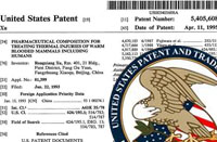
In 2005, we got U.S. patents on the inventions of “Physiological Tissue Repair and Functional Organ Regeneration by Cultivation of Regenerative Stem Cells in Vivo and in Situ'and ‘Composition and Method for Culturing Potentially Regenerative Cells and Functional Tissue-Organs in Vitro.” (US patent numbers 6685971, 6972195, and 6991813). The intellectual property rights on these inventions will be protected for 21 years (Figure 7A-F). It indicates that we have stood in the very front edge of the research and practice in regenerative medicine. After review of our application materials, nine examiners in the U.S. Patent Office stated that they could not imagine how many great changes would take place around the world in the following years by applying our spectacular inventions. Surely our research achievements have brought a great advancement in life science. Over the years, scientists had worked on in vitro organ cloning, and expected the cloned organ could be transplanted into human bodies. Our technologies made transplantation of cloned organs not necessary in many instances, as human organs can be regenerated or renewed at their original sites. Moreover, lives can be extended and diseases can be cured. Some day, medical hospitals may be transformed to health caring centers. Today mankind fights for petroleum; tomorrow people may fight for life regenerative nutrients for survival, health, and longevity. We believe our technologies will eventually change world's economic and even political structures.
IV. Human Lifespan could be Extended
According to the Oxford English Corpus, an English vocabulary database established in 2000, which contains more than one billion words, Time, People, and Year are the top 3 most frequently cited words. This may reflect that people sigh with regret of time and life flying by. Of course, it has been a common dream for people to live a prolonged life.
Currently, there are several hereditary and genetic theories explaining human aging process and longevity. One of them is about chromosome telomere theory. Telomere is a region of high repetitive DNA at the end of liner chromosome that functions as a disposable buffer. Every time chromosomes are replicated, the DNA polymerase complex is incapable of replicating all the way to the end of a chromosome; therefore, a small piece of telomere strand is lost in every cell replication cycle. For that reason, telomeres are used as an indicator for cell aging assessment. Today's average human lifespan is about 72 years. If we use the shrinkage of telomere as an aging indicator, the length of telomere strand is only shortened less than one third of its original length, with two third of telomere left unused, when human beings die in their 70es. Based upon this fact, the maximal lifespan for human tissue cells could be up to 300 years. If human lifespan equals human tissue cell's lifespan, then we can expect maximal human lifespan to be 300 years. This concept might make more sense to Western people since it is not merely a scientific hypothesis to them. The Bible indicated that in the beginning, human being's longevity was at least several hundred years. When men began to multiply on the earth, sins suffused the world. Later, God destroyed the world by the Big Flood. After the Big Flood, Human beings started eating meat. The longevity of human was dramatically reduced after several generations since then.
Thanks to the progress in medicine and overall economic development, the average human lifespan has been extended from 28 years to 72 years in the past two millenniums. In recent 5 years, it is accelerated to 74 years. One of the reasons may be caused by the improvement of dietary habits. However, the maximal human lifespan did not prolong at all. We believe that human lifespan can be prolonged if secondary life cells are promptly and effectively regenerated to take over the responsibilities when primary life cells are injured, declined or dead in the middle of human life, causing the organ systems unable to function. In the rat experiments, which are physiologically representable to human, if adequate regenerative nutrient substances are provided, the secondary life cells can start their regenerative processes when needed. As secondary life cells are activated to massively duplicate, and declined organ functions are accurately renovated, life will be maintained healthy. Thus, we call this process organ life relay.
Unfortunately, the regenerative nutrient substances, i.e. the fundamental materials that secondary life cells rely on for regeneration are not abundantly contained in our daily diets. For this reason, the potential benefits of secondary life cells may not be automatically gained. Now let me give you a briefing on the history of human nutrition study and how we explored and discovered human regenerative nutrient materials in our research.
V. Human Basic Nutrition and Regenerative Nutrient Substances
In fact, human beings don't really know what nutrition they get from their daily diets until approximately two centuries ago. For a long period of time, human beings mainly ate meats. Later on, vegetables and grains were added into their diets. When industrial sugar production was boomed in 16 century, people take sugar as an alternative energy source. In 1842, chemists identified three major categories of nutritional elements including carbohydrates, fats, and proteins in our daily diets. Yet, people had no systematic knowledge of vitamins at that time, but noted that one would get sick if vegetables or fruits were lacking in diets. In 1907, a group of small molecules were identified as vitamins, which was the fourth major category of human nutritional elements. Then minerals and other trace elements were also regarded as vital components to human health. Today people may not recognize the importance of regenerative nutrient substances for their health and longevity. However, I believe people will realize it shortly down the road.
Regenerative nutrient substances were unexpectedly discovered during our explorations on burn treatments. We initially focused on a simpler and better way to treat burn injuries aiming to reduce pain and minimize scarring for our patients. When we noted that extensive deep partial thickness burns were healed unscarred by applying MEBO, we realized there must be a distinctive mechanism causing the physiological recovery of the burns, which we never knew before. From then on, we started immunohistochemical studies on burn wounds during their healing processes. After years of endeavors, we finally found that those actively proliferated histocytes in burn wounds after applying MEBO could be labeled with Keratin 19 monoclonal antibodies, which is a biochemical marker of adult skin stem cells. This result revealed that numerous potential regenerative cells in burnt skin were triggered to be transformed to stem cells and eventually formed new skin and healed the wound. It seamlessly explained our clinical observations in burn patients. People may ask us, how potential regenerative cells got activated to function? We presumed that some unclear substances contained in MEBO might have played a key role in this transformation. By analyzing and testing MEBO constituents, we eventually figured out a group of nutrients attributed to the activation of potential regenerative cells. This is how we discovered regenerative nutrient substances, a source of regenerative energies. Such a small occasion has changed life science, which will never be the same as before and has been driven to an unprecedented era. Our achievements in life science have definitely brought people more excitements.
In summary, our achievements in life science are as follows: (1) we discovered secondary life cells in human tissues, what is ordinary histocytes with regenerative potentials; (2) we discovered regenerative nutrient substances, what can initiate and promote proliferations in secondary life cell; (3) we developed in vitro and in vivo secondary life cell cultivation techniques; (4) we established a new medical system, which is in situ organ regenerative medical system; (5) we introduced a new approach to prevent organ disease and slow down the process of organ aging and degeneration. In general, we have accomplished the organ life relay studies in most human tissues and organs and will continuously make endeavors on extending human lifespan.
VI. In Vitro Tissue Organ Cloning and in situ Organ Regeneration
First, let's take a look of in vitro cloning study in mice small intestinal villi. There are countless villi in the surface of small intestines. They are tiny, finger-like structures that protrude from the wall of the intestines, increasing intestinal absorptive surface area, and providing exceptionally efficient absorption of nutrients in the lumen. However, as the progress of aging process, the functions of intestinal villi tend to gradually decline in human lives after middle of age.
Figure 8A-C showed mice intestinal tissue explants were cultivated in vitro with GIC that contained regenerative nutrient substances (test group) and without GIC (control group). After about ten days of cultivation, fresh and sound new villi were regenerated in test group and explants in the control group entirely died.
Heart muscle cells were considered unable to regenerate in nature. However, when the regenerative nutrient substances were provided to the culture media, heart muscle cells began to multiply. Figure 9 showed that regenerated heart muscle cells connected together and then formed a heart muscle tissue.
We also had successfully cloned pancreatic tissue in vitro. Figure 10A-B showed that the aged and fibrotic pancreatic tissue was renewed after cell cultivation with regenerative substances, and the regenerated tissues could secrete insulin during three months of cultivation period. This result unfolded a promising approach to cure diabetes.
In order to reveal the human cell aging process and figure out if this process might be measurable, rat studies were conducted. The usual lifespan of a male rat is about 2-3 years, or on average with 480 days. Three hundred days old rats which were considered as middle aged rats, were divided into two groups. Test group was fed with additional 10% regenerative nutrient substances to normal recipe and control group was fed with additional 10% water to normal recipe. Both groups were examined in the same living environment. The study lasted for 526 days and the rats in both groups became 826 days old which were considered as very old in rat age. Figure 11-22 showed the histological differences between two groups of study rats. The heart muscle cells in the test group that were fed with regenerative nutrients expressed middle age histo-morphological characteristics, and none fibrosis was observed. In contrast, the control group rats that were not fed with regenerative nutrients demonstrated significant fibrotic changes, which were typical for this age. Similar results were observed in liver, lung, kidney, bone marrow, pancreas, seminiferous tubules, brain, stomach, duodenum, small intestine, and adrenal grand. All these organs in the test groups were in obvious functional conditions. However, organs in control groups became fibrotic and fatty degenerated.
These results have shown that we have made an unbelievable progress in anti-aging research. Long-awaited dreams of longevity are coming true. We believe this achievement could be one of the greatest advancements in life science in 21st century. In the coming five years we will apply these achievements in human lives, and we would like to see more and more people who consume the regenerative nutrient substances stay healthy and energetic.
VII. Anticancer Study of Regenerative Nutrient Substances
In the mutagenesis (cancer-inducing) studies, we noted that normal cells cultivated with regenerative nutrient substances would not be induced to cancer cells. In contrast, normal cells cultured without regenerative substances would be easily induced to cancer cells. This can be interpreted as that the lack of regenerative nutrient substances caused the normal cells being induced to cancer cells. Figure 23 showed that BGC-823 gastric carcinoma cell line was cultivated together with normal bone marrow cells. In those cultures that regenerative nutrient substances were added, cancer cells started dying after 48 hours of cultivation and died out in 5 to 8 days. Meanwhile, the normal bone marrow cells had been proliferated and differentiated dramatically; therefore a new bone marrow tissue was formed in the culture media. On the other hand, in those cultures without regenerative nutrient substances, cancer cells suffused the culture media and normal cells disappeared. For a long time, it was believed that both cancer cells and normal cells required the same nutrients and living environments for the growth. Moreover, the reason that the growth of normal cells was restrained when they were cultured with cancer cells was because the cancer cells grew much faster than normal cells. However, our results completely overturned such belief. We found that normal cells and cancer cells required totally different nutrients and environments to grow. Once the nutrient environment favored normal cells, the normal cells grew and the cancer cells were suppressed to grow.
This hypothesis was further proved by the following studies. Instead of mixing cancer cells with normal cells in cultivation, we cultured cancer cells alone in this study, with or without regenerative nutrient substances respectively. The results showed cancer cells cultured with regenerative nutrient substances stopped proliferation after 48 hours of cultivation, and cancer cells cultured without such nutrients continuously proliferated. Figure 24-26 showed culture results in cancer lines of leukemia, hepatocarcinoma, and ascites sarcoma. Based on these demonstrations, we believe that regenerative nutrient substances played a key role in inhibiting proliferation in cancer cells and promoting the growth in normal cells.
We are currently developing a series of anti-cancer formulas through screening and identifying various nutrient combinations in dozens of cancer cell lines. Those formulas that can enhance normal cell growth and inhibit proliferation in certain cancer cells will be developed into functional foods, in the forms of drinks, tablets, capsules, or even conventional foods, for human consumption. We hope this will help people prevent and treat cancers. In hence, we are very proud to announce that a new effective approach to fight cancer is on its way (Figure 27A-B).
VIII. Applications in Organ Regenerative Remedy
The ultimate goal of scientific research is to utilize them for human well-being. We have held such a vision that is, by using our products, to help people maintain organ functions, prevent diseases, and extend lives, theoretically up to 300 years. In order to fulfill this goal, we have set up an Organ Regenerative Remedy Center in Beijing, and it will be put into use by late of 2007. We hope more branches will be established around the world in a near future. Our center will provide tailor-made health service plans for our clients, ensuring their health inside out.
We aim to accomplish this goal by the means other than chemical drugs, although we look squarely at their capacities, we hold in more esteem for simpler, safer, more natural, and more applicable ways in our practice, which is to provide customized combination of regenerative nutrient substances to maintain healthy functions of organ systems in human beings.
The first service plan in our center is providing customized regenerative nutrient dietary recipe. The procedure of developing the recipe is, first to scrub a sample of cells from the body surface of each client, then incubate these cells to tissue organs in our lab. During the cell cultivations, a series of regenerative nutrient combinations will be tested and the growth of the tissue organ in each combination will be assessed. The best combination will be identified and taken as the most suitable regenerative nutrient combination for that client. Additionally, we will analyze the client's blood sample to determine if the client obtains balanced nutrients from their daily diets. By comparing and evaluating these results, we will recommend an ideal dietary menu to that client and show the client what nutrients are needed for his/her tissues to be effectively regenerated; however, these nutrients may not be obtained adequately from daily diets. We believe such services will help our clients pursue health and longevity.
The second service plan is to provide customized dietary menus for cancer patients. Through in vitro cultures of samples of cancer cells and normal cells obtained from the same patient, we analyze the different nutrient combinations needed by each group of cells. As a result, we identify a nutrient combination that can stop cancer cell growth, meanwhile, promote secondary life cell growth. This is like what is stated in the Chinese proverb “kill two birds with one stone.”
The third service plan is restoring gastrointestinal functions. Since no dietary recipe can help until a functional and robust digestive capacity is restored, we will provide our clients dietary supplemental nutrients to renew their gastrointestinal mucosa for approximately three months before other remedy is initiated.
The last service plan is to promote in situ skin organ regeneration, which helps refresh skin cells by supplementing skin regenerative nutrient substances. With all aforementioned services, we ensure our clients a healthy life inside out. This is our commitment and mission.
Dear fellow experts, due to time constraints, I could not go further in detail of all of our work. As you may know that I am writing a book entitled Science of Second Life in Human Beings, which will be soon published in Switzerland. I believe it will again bring excitement to the world. Regenerative medicine research is our common mission. In conclusion, I would like to quote a well-known phrase from the Russian philosopher, Chernyshevsky, who said, “The future is bright and beautiful, love it! Run to it, embrace it, and try to make it a reality!”
Thank you all.